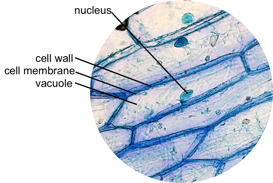
Onion Cell Under Microscope
Finally a light microscope allows you to see the specimen exactly how it is meaning in full color. When a drop of methylene blue is introduced the nucleus is stained which makes it stand out and be clearly seen under the microscope.

Onion Epidermis Under Light Microscope Purple Colored Large
There are two microscope lesson activities in this blog for you to see the nuclei in animal cells and plant cells.

. Plant cells have a cell membrane while animal cells do not. The parts labelled A B and C respectively are. In some of the plants stomata are present on stems and other parts of plants.
You may notice several cells in the scraped material Fig. Draw a labelled. With an electron microscope the image is seen in black and white.
Making up all living material the cell is considered to be the building block of life. Stomata play an important role in gaseous exchange and photosynthesis. List of Microscope Experiments for Kids 1.
It has been designed for biology students at the college and high school level but is useful for medical students physicians science writers and all types of interested people. They control by transpiration rate by opening and closing. Lll G 1 phase Gap 1 lll S phase Synthesis lll G 2 phase Gap 2 G 1 phase corresponds to the interval between mitosis and initiation.
It is easier to see nuclei under a light microscope with staining such as methylene blue. The main onion cell structures are quite easy to observe under medium magnification levels when using a light microscope. Reproducing may be as simple as a single cell dividing to form two daughter cells.
The diagram shows a plant cell as seen under a microscope. Onion skin is an excellent specimen to show plant cells. Onion - A simple layer of onion skin is a great introduction to looking at plant cells.
Show the students the prepared slides before they look at them under the microscope. Turn the coarse focus so that the stage is as close to the objective lens as possible. This work is licensed under a Creative Commons Attribution-NonCommercial-NoDerivs 25 LicenseCreative Commons Attribution-NonCommercial-NoDerivs 25.
When you are ready challenge your knowledge in the testing section to see what you have learned. You should not look. The phases are listed below along with the major events that occur during each phase.
The boundary of the onion cell is the cell membrane covered by another thick covering called the cell wall. The nucleus a component of most eukaryotic cells was identified as the hub of cellular activity 150 years ago. Diagram of onion peel.
Compare the human cells on the left in Figure below and onion cells on the right in. The largest-ever field project investigating evolution began eight years ago with a tweet essentially asking Hey who wants to study clover. Explore topics on usage care terminology and then interact with a fully functional virtual microscope.
The Biology Project Cell Biology Intro to Onion Root Tips Activity Activity Online Onion Root Tips. 2 of 10 STEP 1 -. Because it can be difficult for youngsters to envision such small items have a microscope handy along with prepared slides.
But their cells are very similar. The cell from the Latin word cellula meaning small room is the basic structural and functional unit of life formsEvery cell consists of a cytoplasm enclosed within a membrane which contains many biomolecules such as proteins and nucleic acids. The Biology Project an interactive online resource for learning biology developed at The University of Arizona.
Cells can acquire specified function and carry out various tasks within the cell such as replication DNA repair protein synthesis and. Onion Dissection Look at the Plant Cells. If you looked at cells under a microscope this is what you might see.
Spider Web - Clear nail polish is all you need to see how amazing a spider web really is. Free PDF download of Important Questions with solutions for CBSE Class 8 Science Chapter-8 Cell - Structure and Functions prepared by expert Science teachers from latest edition of CBSENCERT books. The Biology Project is fun richly illustrated and tested on 1000s of students.
Y ou can identify the cell membrane the cytoplasm. Viewed under a light microscope the nucleus appears only as a darker region of the cell but as we increase magnification we find that the nucleus is densely filled with a stew. For animal cells use dyed cheek cells from your own or a students cheek.
An onion a slide and cover slip a cotton bud some food colouring a plate to put the cotton bud on and of course a microscope. The interphase is divided into three further phases. Lesson Description BioNetworks Virtual Microscope is the first fully interactive 3D scope - its a great practice tool to prepare you for working in a science lab.
We can see stomata under the light microscope. Two of the labels are incorrect. Cheek Swab - Take a painless cheek scraping to view the cells in your own body.
Stomata are the tiny openings present on the epidermis of leaves. Harvested cells can provide DNA RNA and protein for the profiling of genomic characteristics gene expression and protein abundance from single-type of cell. The resting phase is the time during which the cell is preparing for division by under going both cell gr owth and DNA r eplication in an or derly manner.
Laser capture microdissection LCM is a technique by which cells of a single-type can be harvested from tissue sections visualized under microscope Chen et al. The cells look elongated similar in appearance- color size and shape- have thick cell walls and a nucleus that is large and circular in shape. Organelles are the functional structures contained inside the cell.
THE TWEET THAT STARTED IT ALL Evolutionary biologists Aleeza Gerstein and Colin Garroway alongside undergraduate student Rebekah Kukurudz in the University of Manitobas Faculty of Science eagerly responded to. Rotate the objective lenses so that the low power eg x10 is in line with the stage. A cell wall B cytoplasm C nucleus.
Every single species is composed of cells including both single celled and multicellular organismsApart from providing shape and structure to an organism the cell performs different functions in order to keep the entire system activeSo the functional structures called organelles inside the cell are. The central dense round body in the centre is called the nucleus. Pond Water - Your favorite pond may be teeming with more life than you think.
Determining time spent in different phases of the cell cycle The life cycle of the cell is typically divided into 5 major phases. Mount a Slide Look at Your Cheek Cells Lesson 3. Although the entire cell appears light blue in color the nucleus at the central part of the cell is much darker which allows it to be identified.
Draw a diagram of Onion peel as observed under a microscope and label its basic components. The nucleus at the central part of the cheek cell contains DNA. The diagram shows a group of onion cells.
The good news is that a computer model can add color for a more realistic view. As you can probably imagine a light microscope is used for more simple study.

Epidermal Onion Cells Under A Microscope Plant Cells Appear Polygonal From The Cell Diagram Plant Cell Diagram Plant Cell

Onion Cells Under A Microscope Requirements Preparation Observation Plant And Animal Cells Animal Cell Plant Cell

Onion Cells Google Images Ilustracoes Felipao

Epidermal Onion Cells Under A Microscope Plant Cells Appear Polygonal From The Cell Diagram Plant Cell Diagram Plant Cell
Comments
Post a Comment